The Truth Is Out There

Posts tagged ‘virology’

Flu Vaccines Contain RNA That Trigger Positive PCR Test Results: ‘Journal of Medical Microbiology’


Is the “chilling” rise in flu cases nationwide attributable to PCR tests detecting vaccine RNA, not wild virus?

Mainstream news outlets are broadcasting that there is a “chilling” rise in flu cases, with Colorado, Louisiana, and New York experiencing the “fastest increases in influenza cases.”

However, the rise in cases follows flu vaccination campaigns in those states, which raises questions about vaccine efficacy.

But it also raises questions about whether the vaccinations themselves are contributing to the increasing case numbers.

For example, the New Orleans Health Department (NOHD) launched a flu vaccination campaign in early October.

NYC Health Department similarly launched an October push urging all residents 6 months and older to get flu shots.

The Colorado Department of Public Health and Environment’s (CDPHE) influenza webpage was updated the same month to promote flu vaccination.

These campaigns are meant to increase flu vaccine uptake.

Now there’s a rise in influenza cases, which are counted using positive PCR test results.


However, a March 2012 Journal of Medical Microbiology publication confirms the presence of residual viral RNA (genomic RNA—which PCR tests look for—from the influenza viruses used in vaccine production) in inactivated split-virus seasonal influenza vaccines.

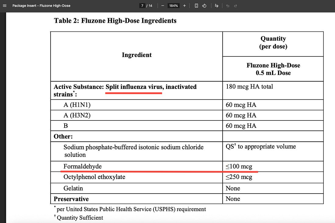
One of the most popular injectable flu vaccines in the U.S., the formaldehyde-containing ‘Fluzone High-Dose,’ is an inactivated split-virus vaccine.

The 2012 study directly tested two 2010 trivalent inactivated vaccines (egg-based, similar in type to Fluzone) and detected high quantities of influenza A and B viral RNA using real-time RT-PCR on the vaccine liquid itself.

This RNA was stable, remaining detectable for at least 66 days after opening the vials.

Sequencing confirmed it included genetic components matching vaccine strains.

The study abstract reads:

False-positive PCR results usually occur as a consequence of specimen-to-specimen or amplicon-to-specimen contamination within the laboratory. Evidence of contamination at time of specimen collection linked to influenza vaccine administration in the same location as influenza sampling is described. Clinical, circumstantial and laboratory evidence was gathered for each of five cases of influenza-like illness (ILI) with unusual patterns of PCR reactivity for seasonal H1N1, H3N2, H1N1 (2009) and influenza B viruses. Two 2010 trivalent influenza vaccines and environmental swabs of a hospital influenza vaccination room were also tested for influenza RNA. Sequencing of influenza A matrix (M) gene amplicons from the five cases and vaccines was undertaken. Four 2009 general practitioner (GP) specimens were seasonal H1N1, H3N2 and influenza B PCR positive. One 2010 GP specimen was H1N1 (2009), H3N2 and influenza B positive. PCR of 2010 trivalent vaccines showed high loads of detectable influenza A and B RNA. Sequencing of the five specimens and vaccines showed greatest homology with the M gene sequence of Influenza A/Puerto Rico/8/1934 H1N1 virus (used in generation of influenza vaccine strains). Environmental swabs had detectable influenza A and B RNA. RNA detection studies demonstrated vaccine RNA still detectable for at least 66 days. Administration of influenza vaccines and clinical sampling in the same room resulted in the contamination with vaccine strains of surveillance swabs collected from patients with ILI. Vaccine contamination should therefore be considered, particularly where multiple influenza virus RNA PCR positive signals (e.g. H1N1, H3N2 and influenza B) are detected in the same specimen.

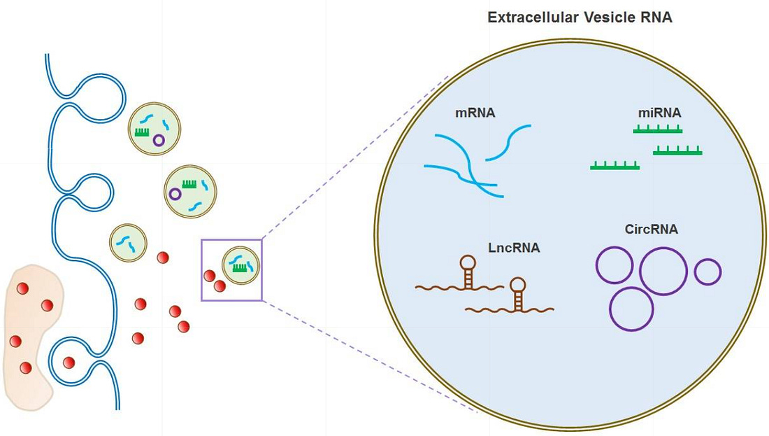
The human body’s own extracellular vesicles (EVs) (including exosomes) naturally carry and transfer various RNAs (herehereherehere) through bodily fluids such as blood, saliva, urine, nasal mucus, and cerebrospinal fluid.

These are the exact substances PCR tests are applied to.

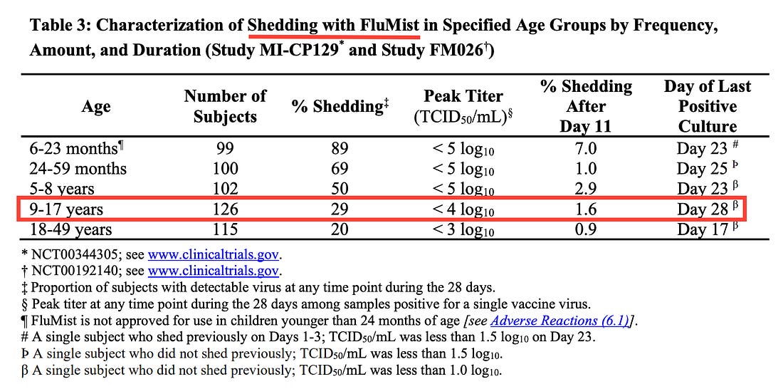
Another popular flu vaccine is FluMist, whose FDA package insert directly confirms uses a live virus that can be shed from bodily fluids for at least 28 days after vaccination, detectable with PCR tests.

All together, this data raises logical questions:

  • Are PCR tests detecting vaccine virus RNA, not wild virus RNA?
  • Is the nationwide rise in flu-positive PCR tests attributable, at least in part, to the detection of vaccine material?
  • Why haven’t the CDC or vaccine manufacturers directly tested this?

32% of COVID ‘Spike Protein’ Matches Human Genes—Suggesting Either Contamination or Intentional Chimeric Design: BLASTp Analysis


One-third of the so-called SARS-CoV-2 spike protein was assembled from human genetic material, not viral RNA—raising urgent questions about the origins of the sequence used in mRNA vaccines.

3D print of a spike protein on the surface of SARS-CoV-2—also known as 2019-nCoV, the virus that causes COVID-19. Spike proteins cover the surface of SARS-CoV-2 and enable the virus to enter and infect human cells. For more information, visit the NIH 3D Print Exchange at 3dprint.nih.gov. Credit: NIH/Wikimedia Commons under the Creative Commons Attribution 2.0 Generic license. Image saturation, vibrace, color, and temperature have been altered in Canva Pro.

The most critical sequence in modern medical history—the “spike protein” of SARS-CoV-2—may never have existed in nature at all.

As a new research paper explains, A 32% Human-Derived Mosaic in the In Silico-Assembled SARS-CoV-2 Spike Protein: Accidental Contaminant Misincorporation or Intentional Functional Chimeric Design?, the Wuhan “spike” was never isolated from a virus.

It was digitally stitched together—in silico (in a computer)—from fragments of RNA found in the lung fluid of one patient in China by Chinese researchers in early January 2020.

The genetic information was rapidly disseminated through databases such as GenBank and GISAID.

The foundational publication by Wu et al. in Nature in February 2020 represented the first peer-reviewed article presenting the full genome sequence of the novel coronavirus (SARS-CoV-2), including its spike protein sequence.

That synthetic model then became the blueprint for the Pfizer and Moderna mRNA vaccines injected into over five billion people worldwide.

This raises a critical question: if the Wuhan team’s “virus” was assembled from a sick man’s lung fluid, why does its defining spike protein contain extensive human genetic material—was this simply contamination from the patient’s own RNA, or evidence that the sequence was artificially constructed using human genes?

To identify the human components of that digital construct, I used the NCBI BLASTp tool—a government-hosted bioinformatics search engine that compares protein sequences against the entire global database of known organisms—to systematically test the Wuhan spike protein for matches to human proteins, revealing extensive alignments to human endogenous retroviruses and cellular genes that are absent in any bat or pangolin coronavirus.

The full research article, along with all six NCBI BLASTp run raw data files and their reproducible Request IDs, has been publicly archived on Zenodo (DOI 10.5281/zenodo.17583428), ensuring complete transparency and independent verification of every alignment reported.

You can also read and download the research article below:

A 32% Human Derived Mosaic In The In Sil…724KB ∙ PDF file
Download

Key Findings

  • 32% of the spike protein—416 amino acids—matches human genetic material.
    The overlaps include sequences from human endogenous retroviruses (HERV-K, HERV-H, HERV-W) and cellular proteins linked to immune modulation, fusion, and intracellular trafficking.
  • No comparable overlaps exist in bat or pangolin coronaviruses.
    These human alignments appear only in the SARS-CoV-2 spike, not in its supposed animal precursors.
  • Six independent NCBI BLASTp runs confirm the findings.
    Each run produced reproducible, statistically significant human alignments—with probabilities of random occurrence as low as one in 10²⁰.
  • Critical overlaps occur in known functional domains:
    • HERV-K envelope homology in the S2 fusion region, which controls cell-to-cell syncytia.
    • HERV-H alignment at the furin cleavage site, already linked to a patented human gene (MSH3).
    • HERV-W (MSRV) match in the N-terminal domain, associated with neuroinflammation.
    • Additional matches with human lysosomal, mitochondrial, and zinc-finger proteins that govern energy metabolism and DNA regulation.

Why It Matters

If one-third of the spike’s code came from human sources, two explanations remain:

  1. Accidental misassembly—contamination from human RNA in the original Wuhan sample (BALF), which was never purified before computational assembly; or
  2. Intentional inclusion—deliberate use of human sequences to enhance infectivity, persistence, or immune modulation.

Both possibilities challenge the official story that the SARS-CoV-2 genome was a “naturally emerging” virus.

Independent Validation

Multiple clinical studies now confirm that these same HERV sequences are biologically active in COVID-19 patients:

  • Petrone et al., 2023: HERV-K and HERV-W upregulated in nasal mucosa; expression levels predict hospitalization.
  • Temerozo et al., 2022: HERV-K found in lung aspirates of deceased ICU patients.
  • Guo et al., 2022: HERV activation triggers interferon and inflammation via cGAS-STING.
  • Balestrieri et al., 2023: HERV-W linked to pediatric MIS-C and Kawasaki-like syndromes.
  • Wang et al., 2023: HERV-K expression correlates with pulmonary hypertension.
  • Wu et al., 2025: SARS-CoV-2 directly transactivates HERV-K, producing retrovirus-like particles that drive senescence and neurodegeneration.

Together, these findings corroborate that the “human” portions of the spike aren’t computational noise—they’re functionally active biological components.

Bottom Line

The official reference spike (YP_009724390.1)—used worldwide in vaccine development—is a digital, computationally assembled sequence derived from patient RNA rather than from a purified viral protein.

This sequence contains hundreds of human gene fragments precisely placed within key functional domains impacting viral fusion, immune evasion, and inflammation pathways.

While standard re-assembly of the original raw sequencing data (SRR10971381) excluding human reads has been performed, no published study has conducted an independent, viral-only de novo assembly focusing specifically on the spike region to test for potential human contaminant incorporation.

Thus, the origin of this 32% human mosaic—whether due to accidental laboratory contamination or intentional chimeric engineering—remains unresolved and requires targeted re-analysis.